Here is a selection of old anatomical illustrations that provide a unique perspective on the evolution of medical knowledge in Japan during the Edo period (1603-1868).
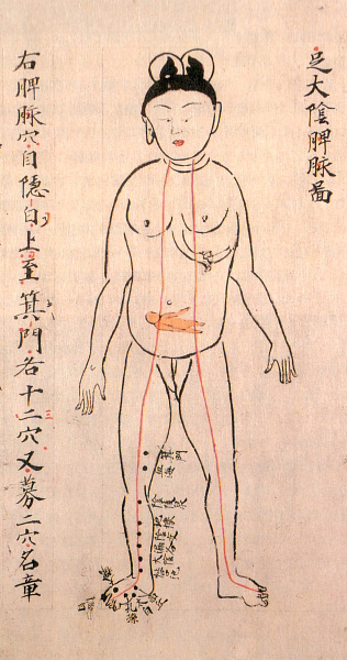
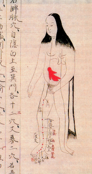
Pregnancy illustrations, circa 1860
These pregnancy illustrations are from a copy of Ishinhō, the oldest existing medical book in Japan. Originally written by Yasuyori Tanba in 982 A.D., the 30-volume work describes a variety of diseases and their treatment. Much of the knowledge presented in the book originated from China. The illustrations shown here are from a copy of the book that dates to about 1860.
* * * * *
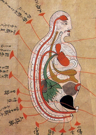
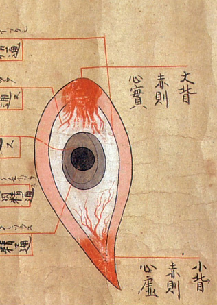
Anatomical illustrations, late 17th century [+]
These illustrations are from a late 17th-century document based on the work of Majima Seigan, a 14th-century monk-turned-doctor. According to legend, Seigan had a powerful dream one night that the Buddha would bless him with knowledge to heal eye diseases. The following morning, next to a Buddha statue at the temple, Seigan found a mysterious book packed with medical information. The book allegedly enabled Seigan to become a great eye doctor, and his work contributed greatly to the development of ophthalmology in Japan in the 16th and 17th centuries.
* * * * *
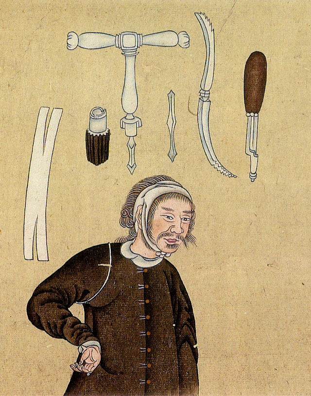
Trepanning instruments, circa 1790 [+]
These illustrations are from a book on European medicine introduced to Japan via the Dutch trading post at Nagasaki. Pictured here are various trepanning tools used to bore holes in the skull as a form of medical treatment.
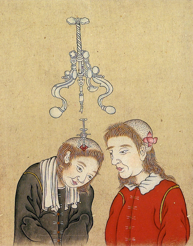
Trepanning instruments, circa 1790 [+]
The book was written by Kōgyū Yoshio, a top official interpreter of Dutch who became a noted medical practitioner and made significant contributions to the development of Western medicine in Japan.
* * * * *
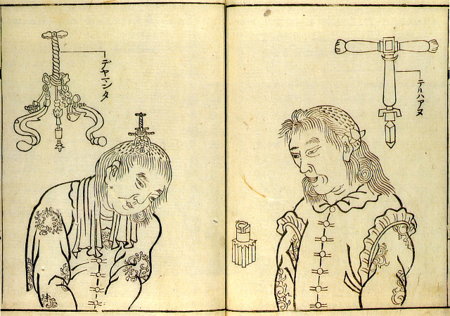
Trepanning instruments, 1769 [+]
These illustrations of trepanning instruments appeared in an earlier book on the subject.
* * * * *
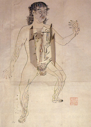
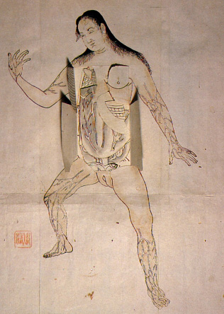
Anatomical illustrations (artist/date unknown) [+]
These anatomical illustrations are based on those found in Pinax Microcosmographicus, a book by German anatomist Johann Remmelin (1583-1632) that entered Japan via the Dutch trading post at Nagasaki.
* * * * *
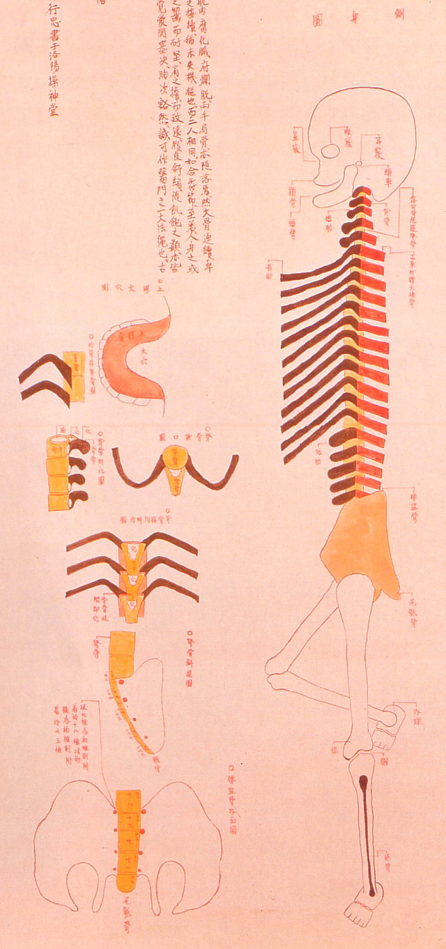
Human skeleton, 1732
These illustrations -- created in 1732 for an article published in 1741 by an ophthalmologist in Kyōto named Toshuku Negoro -- show the skeletal remains of two criminals that had been burned at the stake.
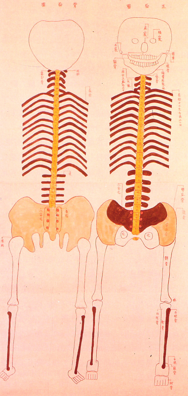
Human skeleton, 1732
This document is thought to have inspired physician Tōyō Yamawaki to conduct Japan's first recorded human dissection.
* * * * *
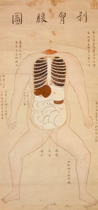
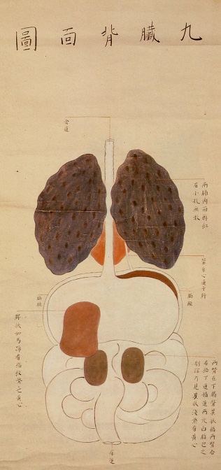
Japan's first recorded human dissection, 1754
These illustrations are from a 1754 edition of a book entitled Zōzu, which documented the first human dissection in Japan, performed by Tōyō Yamawaki in 1750. Although human dissection had previously been prohibited in Japan, authorities granted Yamawaki permission to cut up the body of an executed criminal in the name of science.
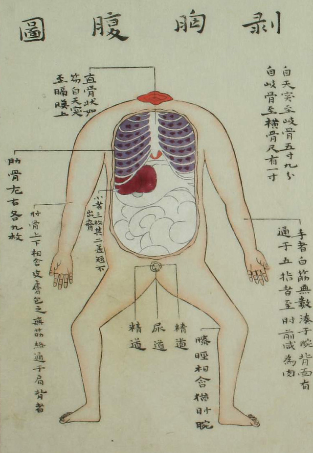
Illustration from 1759 edition of Zōzu
The actual carving was done by a hired assistant, as it was still considered taboo for certain classes of people to handle human remains.
* * * * *
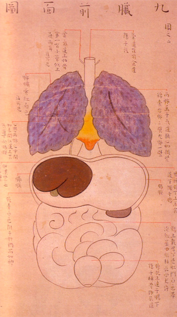
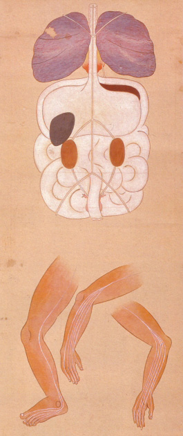
Japan's second human dissection, 1758 // First human female dissection, 1759
In 1758, a student of Tōyō Yamawaki's named Kōan Kuriyama performed Japan's second human dissection (see illustration on left). The following year, Kuriyama produced a written record of Japan's first dissection of a human female (see illustration on right). In addition to providing Japan with its first real peek at the female anatomy, this dissection was the first in which the carving was performed by a doctor. In previous dissections, the cutting work was done by hired assistants due to taboos associated with handling human remains.
* * * * *
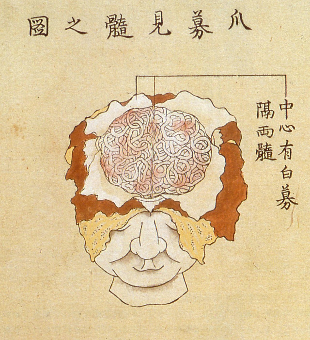
Kaishihen (Dissection Notes), 1772
Japan's fifth human dissection -- and the first to examine the human brain -- was documented in a 1772 book by Shinnin Kawaguchi, entitled Kaishihen (Dissection Notes). The dissection was performed in 1770 on two cadavers and a head received from an execution ground in Kyōto.
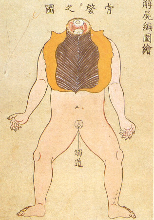
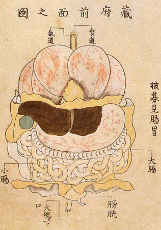
Kaishihen (Dissection Notes), 1772
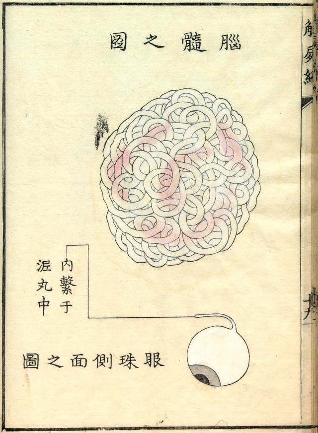
Kaishihen (Dissection Notes), 1772
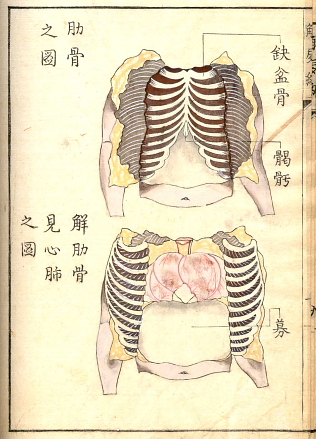
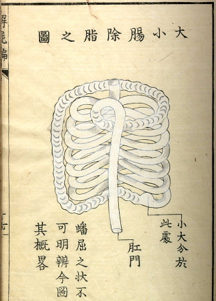
Kaishihen (Dissection Notes), 1772
* * * * *
Tōmon Yamawaki, son of Tōyō Yamawaki, followed in his father's footsteps and performed three human dissections.
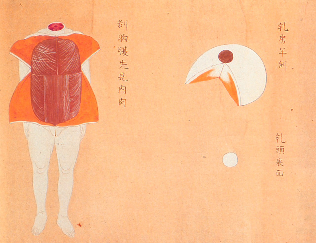
Female dissection, 1774
He conducted his first one in 1771 on the body of a 34-year-old female executed criminal. The document, entitled Gyokusai Zōzu, was published in 1774.
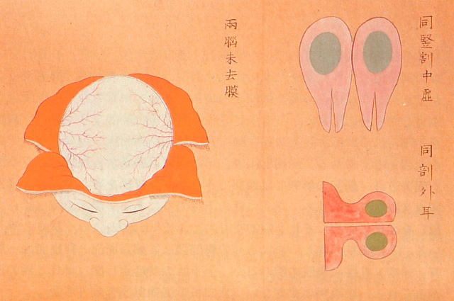
Female dissection, 1774
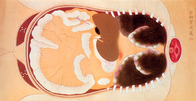
Female dissection, 1774
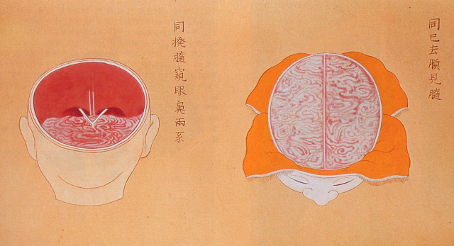
Female dissection, 1774
* * * * *
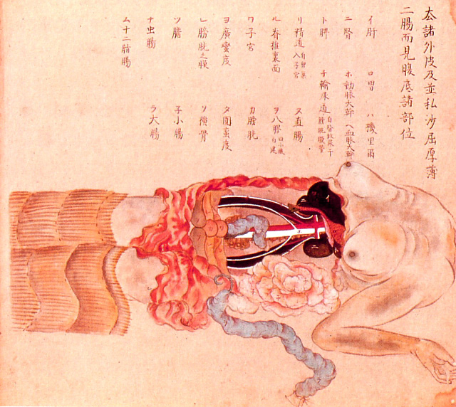
Female dissection, 1800
These illustrations are from a book by Bunken Kagami (1755-1819) that documents the dissection of a body belonging to a female criminal executed in 1800.
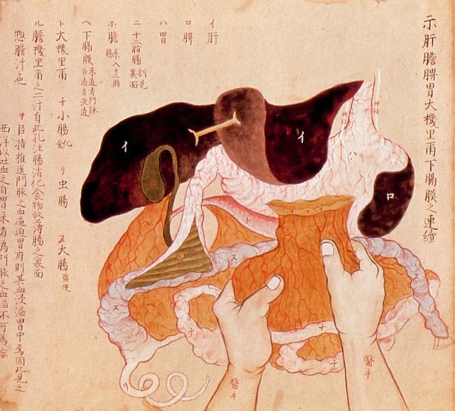
Female dissection, 1800
* * * * *
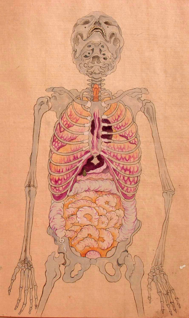
Human anatomy (date unknown)
This anatomical illustration is from the book Kanshin Biyō, by Bunken Kagami.
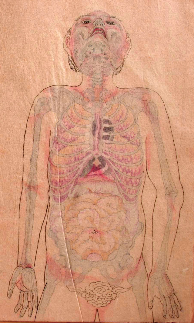
Human anatomy (date unknown)
In this image, a sheet of transparent paper showing the outline of the body is placed over the anatomical illustration.
* * * * *
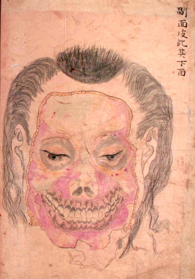
Seyakuin Kainan Taizōzu (circa 1798)
These illustrations are from the book entitled Seyakuin Kainan Taizōzu, which documents the dissection of a 34-year-old criminal executed in 1798. The dissection team included the physicians Kanzen Mikumo, Ranshū Yoshimura, and Genshun Koishi.
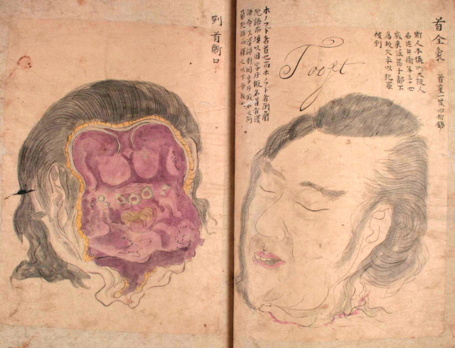
Seyakuin Kainan Taizōzu (circa 1798)
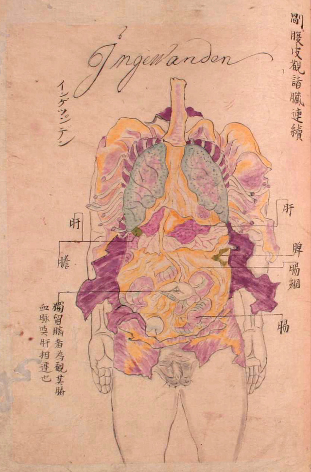
Seyakuin Kainan Taizōzu (circa 1798)
* * * * *
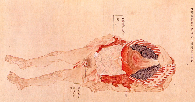
Dissection, 1783 [+]
This illustration is from a book by Genshun Koishi on the dissection of a 40-year-old male criminal executed in Kyōto in 1783.
* * * * *
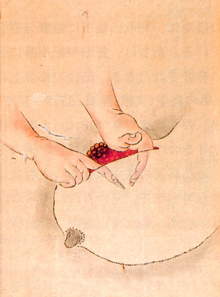
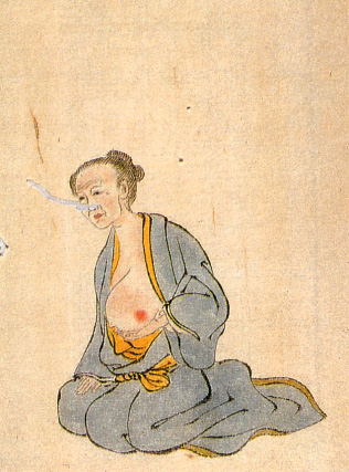
Breast cancer treatment, 1809
These illustrations are from an 1809 book documenting various surgeries performed by Seishū Hanaoka for the treatment of breast cancer. The illustrations here depict the treatment for a 60-year-old female patient.
* * * * *
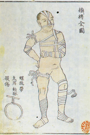
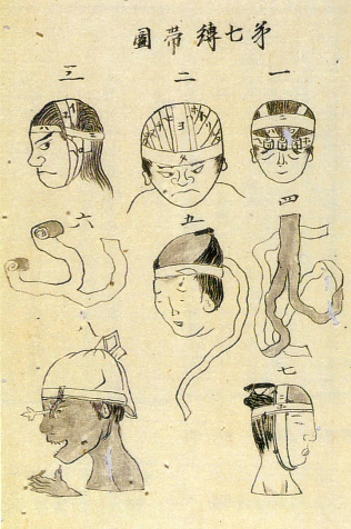
Bandage instructions from two medical encyclopedias, 1813
* * * * *
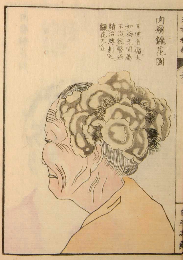
Yōka Hiroku (Confidential Notes on the Treatment of Skin Growths), 1847
These illustrations are from the 1847 book Yōka Hiroku (Confidential Notes on the Treatment of Skin Growths) by surgeon Sōken Honma (1804-1872).
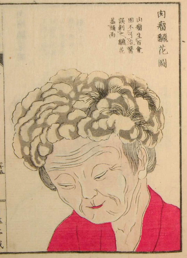
Yōka Hiroku (Confidential Notes on the Treatment of Skin Growths), 1847
* * * * *
The following illustrations are from the 1859 book Zoku Yōka Hiroku (Sequel to Confidential Notes on the Treatment of Skin Growths), an 1859 book by Sei Kawamata that presented the teachings of surgeon Sōken Honma.
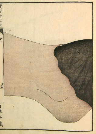
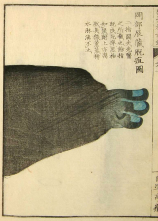
Zoku Yōka Hiroku (Sequel to Confidential Notes on the Treatment of Skin Growths), 1859
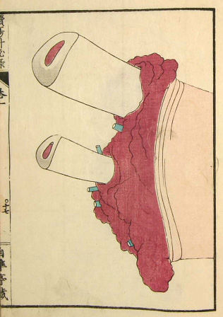
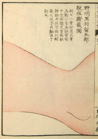
Zoku Yōka Hiroku (Sequel to Confidential Notes on the Treatment of Skin Growths), 1859
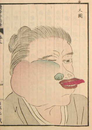
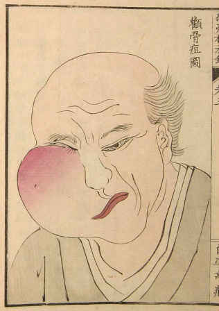
Zoku Yōka Hiroku (Sequel to Confidential Notes on the Treatment of Skin Growths), 1859
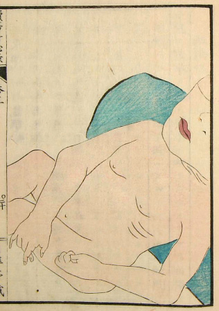
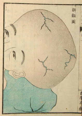
Zoku Yōka Hiroku (Sequel to Confidential Notes on the Treatment of Skin Growths), 1859
[Source: Nihon Iryō Bunkashi (History of Japanese Medical Culture), Shibunkaku Publishing, 1989]

MsJulie
Thank you for taking the trouble to compile this. Very fascinating. Makes me grateful I live now, though I wonder what people will think of our level of knowledge.
[]jessie
This is a wonderful look into both the arts and sciences of that time. If you think about their level of understanding of the functionality of the organs, the artists did a fantastic job depicting them in both color and placement. I am especially interested in the poses of the subjects. They were unafraid to draw fat, skin, or nipples. Also, the stances of the skeletal drawings were very interesting to me because the heads (or skulls) were angled at a difficult angle to draw, where we could see up into the crevices. I really like that the cultural differences are clear, never mind the foreign text.
[]henk
Amazing drawings. "Seyakuin Kainan TaizÅzu (circa 1798)" - it says "ingewanden" in the top of the drawing. It's Dutch for "insides". This particular drawing has to be made in the complete south of Japan.
[]D
興味深ã„ã§ã™ã€‚ã“ã†ã„ã†ã®ã©ã£ã‹ã‚‰æŽ¢ã—ã¦ãã‚‹ã‚“ã§ã™ã‹ï¼Ÿ
[]Graham
amazing. i would love to buy prints of some of these.
[]Duck
The progression is very cool. The oldest images are so vague, which makes sense as they are guess work, more or less, while the most recent (150 years old but still) are far more accurate. The differences in the the depictions of the skull and intestines show the progress so well. It just goes to show the importance of autopsy, dissection, and experimentation. It might not sound pleasant but it's the only way medicine could (and can) evolve.
[]dole
I smell a cool new Trauma Center game!
[]Jappleng
I'll take the worst surgeon that exists today before them lol! I feel pretty queezy now, thanks a lot! :(
[]Fi
What a wonderful find! I've seen plenty of early Western-style anatomy pictures and I find it fascinating to see these which have such a totally different emphasis on which details were chosen to be recorded.
[]Matthew Meyer
These are so gorgeous!! Like Fi said above, I've seen countless anatomical drawings from Western painters dating back so many centuries, but I'd never seen such gorgeous Japanese medical drawings before.
More artwork like this please! :-)
[]AdelaideBen
Bizarre - and yet rivetting. It's like watching the drawings of a small child, growing up to maturity, right before your eyes It's somewhat scary to think of the lack of knowledge that existed... and how much we take for granted now.
[]PBL
Maravillosos.
[]VentingPost.com
What I find hard to understand is how did "man" NOT understand what they drew does NOT look like what they're drawing. So hard for me to accept.
I mean they knew well enough to make the finest blades in the world and accurately create straight and true weapons but not the human body?
However, 200yrs before in Leonardo's time
http://en.wikipedia.org/wiki/File:Da_Vinci_Studies_of_Embryos_Luc_Viatour.jpg
was able to depict in correct portions and position...
Oh not to mention even in the Edo period, they created Samurai houses without nails! All absolute perfectly interlocking joints. Even asof today one still can't do that without the aid of a cadcam/machine.
[]James Del Nero
where did you find this information?
[]Kristina
Do you know where I can get a copy of this book:
[][Source: Nihon Iryō Bunkashi (History of Japanese Medical Culture), Shibunkaku Publishing, 1989]? Or an archive/rare books library that has a copy?
rocio
THIS IS AMAZING, thank you so much for sharing! Just wanted to say it.
[]Malcolm Clements
Eye diseases can be prevented through food supplements and also making sure that your eye gets a good rest after eye intensive tasks like working on a computer for long periods of time. ,:*:"
Many thanks http://healthmedicinelab.com/appendicitis-symptoms/
[]